



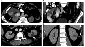
Source : AJKD , Computed tomography images from patients (A, B) 1 and (C-D) 2. (A) Axial and (B) coronal direction in the portal venous phase shows multiple perfusion defects (arrows) in the kidney allograft, most pronounced in the upper pole. (C) Axial and (D) coronal direction in the portal venous phase indicates a wedge-shaped perfusion defect dorsolateral in the interpolar area of the left kidney










Leave a Reply